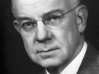
Maurice Hugh Frederick Wilkins (1916-2004)
On December 15, 1916, New Zealand-born British physicist, molecular biologist, and Nobel Laureate Maurice Wilkins was born. Wilkins’ research contributed to the scientific understanding of phosphorescence, isotope separation, optical microscopy and X-ray diffraction, and to the development of radar. He is best known for his work at King’s College London on the structure of DNA.
“It is essential for genetic material to be able to make exact copies of itself; otherwise growth would produce disorder, life could not originate, and favourable forms would not be perpetuated by natural selection.”
― Maurice Wilkins
Youth and Education
Maurice Wilkins was born in Pongaroa, north Wairarapa, New Zealand where his father, Edgar Henry Wilkins was a medical doctor. Originally his family had come from Dublin, but at age 6, Maurice’s family moved to Birmingham, England, where, he attended Wylde Green College and then went to King Edward’s School from 1929 to 1934. Wilkins went to St John’s College, Cambridge in 1935 to study physics, within the Natural Sciences Tripos, and received a B.A. Wilkins became a Ph.D. student of John T. Randall at Birmingham University, where he studied the luminescence of solids. In 1945, they published three papers in the Proceedings of the Royal Society on phosphorescence and electron traps. Wilkins received his Ph.D. for this work, being mainly on a study of thermal stability of trapped electrons in phosphors, and on the theory of phosphorescence, in terms of electron traps with continuous distribution of trap depths.[1] Next he worked under M. L. E. Oliphant on mass spectrograph separation of uranium isotopes for use in bombs and, shortly after, moved with others from Birmingham to the Manhattan Project in Berkeley, California, where these studies continued.[1]
Academic Career
Meanwhile, Randall had been appointed to the Chair of Physics at the University of St Andrews, where he appointed Wilkins as Assistant Lecturer in his department at the University of St Andrews in 1945. Randall was negotiating with the Medical Research Council (MRC) to set up a laboratory to apply the experimental methods of physics to problems of biology. The combination of these disciplines as biophysics was a novel idea. The MRC told Randall that this had to be done in another university. In 1946 Randall was appointed Wheatstone Professor of Physics, in charge of the entire Physics department at King’s College, London, with the funding to set up a Biophysics Unit. He brought Wilkins with him, as Assistant Director of the unit. They appointed a team of scientists trained in both the physical and biological sciences.
Ultrasonics, Reflecting, and Inference Microscopes
He was first concerned with genetic effects of ultrasonics; after one or two years, he changed his research to development of reflecting microscopes for ultraviolet microspectrophotometric study of nucleic acids in cells. He also studied the orientation of purines and pyrimidines in tobacco mosaic virus and in nucleic acids, by measuring the ultraviolet dichroism of oriented specimens, and he studied, with the visible-light polarizing microscope, the arrangement of virus particles in crystals of TMV and measured dry mass in cells with interference microscopes.[1]
X-Ray Diffraction
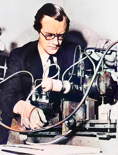
At King’s College Wilkins pursued, among other things, X-ray diffraction work on ram sperm and DNA that had been obtained from calf thymus by the Swiss scientist Rudolf Signer. The DNA from Signer’s lab was much more intact than the DNA which had previously been isolated. Wilkins discovered that it was possible to produce thin threads from this concentrated DNA solution that contained highly ordered arrays of DNA suitable for the production of X-ray diffraction patterns. Using a carefully bundled group of these DNA threads and keeping them hydrated, Wilkins and a graduate student Raymond Gosling obtained X-ray photographs of DNA that showed that the long, thin DNA molecule in the sample from Signer had a regular, crystal-like structure in these threads.
The Hunt for the DNA Structure
It was one of the X-ray diffraction photographs taken in 1950, shown at a meeting in Naples a year later, that sparked James Watson’s interest in DNA. At that time Wilkins also introduced Francis Crick to the importance of DNA [2]. Crick advised him to work on proteins with improved X-ray equipment and a new microcamera. He also suggested to Randall that the soon-to-be-appointed Rosalind Franklin should be reassigned from work on protein solutions to join the DNA effort. Late in 1950, Randall wrote to Franklin to inform her that rather than work on protein, she should take advantage of Wilkins’s preliminary work and that she should do X-ray studies of DNA fibers made from Signer’s samples of DNA.[3]
By November 1951, Wilkins had evidence that DNA in cells as well as purified DNA had a helical structure. Alex Stokes had solved the basic mathematics of helical diffraction theory and thought that Wilkins’s X-ray diffraction data indicated a helical structure in DNA. Wilkins met with Watson and Crick and told them about his results. This information from Wilkins, along with additional information gained by Watson when he heard Franklin talk about her research during a King’s College research meeting, stimulated Watson and Crick to create their first molecular model of DNA. Franklin was able to generate newer data on DNA amidst a swirl of conflict between colleagues at different institutions. Wilkins later showed a DNA photo taken by Franklin’s graduate assistant on a machine she had refined to competing scientist James D. Watson. He and Francis Crick used the image, along with additional information and their own scientific knowledge, to support their theory of DNA’s double helix structure, which was published in 1953.[4] If Wilkins and Franklin had cooperated better, they might have been the first to discover DNA’s structure.[5]
Nobel Prize and Later Years
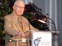
Following the initial 1953 series of publications on the double helix structure of DNA, Wilkins continued research as leader of a team that performed a range of meticulous experiments to establish the helical model as valid among different biological species, as well as in living systems, to establish the universality of the double helix structure. He became deputy director of the MRC Biophysics Unit at King’s in 1955, and succeeded Randall as director of the unit from 1970 to 1972. In 1962 Wilkins shared the Nobel Prize in Physiology or Medicine with Watson and Crick “for their discoveries concerning the molecular structure of nucleic acids and its significance for information transfer in living material“. In 1969, Wilkins became the founding President of the British Society for Social Responsibility in Science.
Maurice Wilkins died in Blackheath, London, on 5 October 2004, at age 87.
Erik Lindhal, Lecture 01, concept 02: From data and models to DNA structure, [8]
References and Further Reading:
- [1] Maurice Wilkins – Biographical, at Nobelprize.org
- [2] Crick and Watson decipher the DNA, SciHi blog, February 28, 2013.
- [3] Rosalind Franklin and the Beauty of the DNA Structure, SciHi blog, July 27, 2014.
- [4] Maurice Wilkins, at Biography.com
- [5] Maurice Wilkins: Behind the Scenes of DNA, at nature.com, Essentials of Genetics, Unit 1.3
- [6] The first American newspaper coverage of the discovery of the DNA structure: Saturday, 13 June 1953 The New York Times
- [7] Maurice Wilkins at Wikidata
- [8] Erik Lindhal, Lecture 01, concept 02: From data and models to DNA structure, Molecular Biophysics, Erik Lindahl @ youtube
- [9] Arnott, S.; Kibble, T. W. B.; Shallice, T. (2006). “Maurice Hugh Frederick Wilkins. 15 December 1916 – 5 October 2004: Elected FRS 1959”. Biographical Memoirs of Fellows of the Royal Society. London: Royal Society. 52: 455–478.
- [10] Witkowski J (2019). “The forgotten scientists who paved the way to the double helix”. Nature. 568 (7752): 308–309.
- [11] Randall, J. T.; Wilkins, M. H. F. (1945). “The Phosphorescence of Various Solids”. Proceedings of the Royal Society A: Mathematical, Physical and Engineering Sciences. 184 (999): 347–364.
- [12] Maurice Wilkins, The Third Man of the Double Helix: The Autobiography of Maurice Wilkins. Oxford University Press
- [13] Robert Olby; The Path to The Double Helix: Discovery of DNA; first published in October 1974 by MacMillan
- [14] Olby, R. (2003). “Quiet debut for the double helix”. Nature. 421 (6921): 402–405.
- [15] Timeline of Maurice Wilkins at Wikidata